Home
/ Head Digestive System Model Labeled - Alila Medical Media Digestive Organs And Bile Ducts Labeled Diagram Medical Illustration, Torso model with key organs of the digestive system.
Head Digestive System Model Labeled - Alila Medical Media Digestive Organs And Bile Ducts Labeled Diagram Medical Illustration, Torso model with key organs of the digestive system.
Head Digestive System Model Labeled - Alila Medical Media Digestive Organs And Bile Ducts Labeled Diagram Medical Illustration, Torso model with key organs of the digestive system.. The digestive system is made up of the digestive tract and other organs that help the body break down and absorb food. Learn vocabulary, terms and more with flashcards, games and other study tools. Digestive system mounted on baseboard for easy a smart label with nfc technology and a qr code can be located on each original 3b scientific anatomy model. They should include a description of what happens at each stage in the process along with the label. The human digestive system consists of the gastrointestinal tract plus the accessory organs of digestion (the tongue, salivary glands, pancreas, liver, and gallbladder).
Find out about how it works and some related conditions. This worksheet was designed for anatomy students to practice labeling the organs of the digestive system. To unlock these benefits, simply scan the label located on your model and register online. Digestive systems take many forms. Download media please credit each image as:
Chapter 24 Digestive System Flashcards Quizlet from quizlet.com Your digestive system is a sophisticated machine that absorbs the food you eat and transforms it into energy and nutrients. Zoom out so that the entire model is visible. → nose → mouth cavity and pharynx → esophagus → gi tract → liver with gall bladder → pancreas → spleen. 500 x 500 jpeg 34 кб. In this activity, students will label a model of the digestive system. Torso model with key organs of the digestive system. Please like, share and subscribe!!! The alimentary canal is made up.
Find out about how it works and some related conditions.
Zoom out so that the entire model is visible. ● describe the path you (the food) take as you travel. Start studying digestive system model/label. Human digestive system, system used in the human body for the process of digestion. Human male body digestive system intestine colon stomach liver pancreas man head face character organ organs textured anatomy. Label the digestive organs listed in this exercise on the model pictures on the following pages. Upper digestive system go to the views menu and select 1. 960 x 720 jpeg 59 кб. To unlock these benefits, simply scan the label located on your model and register online. Our tips from experts and exam survivors will help you through. Teach patients and students the location and function of gastrointestinal organs. This high quality digestive system is a great teaching tool for any anatomy lesson or doctor's office. Digestion is the breakdown of carbohydrates, proteins and fats into small muscular organ where digestion continues.
This worksheet was designed for anatomy students to practice labeling the organs of the digestive system. Your digestive system breaks down the food you eat into nutrients such as carbohydrates, fats and proteins. The digestive system is made up of the digestive tract and other organs that help the body break down and absorb food. Your students and patients will be able to understand their lesson or appointment fully with our anatomical replicas of the human. In most vertebrates, digestion is a multistage process in the digestive system, starting from ingestion of raw materials, most often other organisms.
The Human Digestive System Organs Functions And Diagram from microbenotes.com The tissue in this tract can be divided into four layers it contains blood vessels, nerve endings, lymphatic vessels, and cells of the immune system. 960 x 720 jpeg 59 кб. The human digestive system consists of the gastrointestinal tract plus the accessory organs of digestion (the tongue, salivary glands, pancreas, liver, and gallbladder). Your digestive system breaks down the food you eat into nutrients such as carbohydrates, fats and proteins. Our tips from experts and exam survivors will help you through. ● describe the path you (the food) take as you travel. Teach patients and students the location and function of gastrointestinal organs. The human digestive system consists primarily of the digestive tract, or the series of structures and organs through which food and liquids pass during their processing into forms absorbable into the bloodstream.
You can use this digestive system worksheet with your ks2 science and biology class to test their knowledge of human anatomy and the different parts of our digestive system.
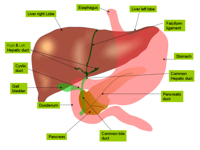
To scaffold this activity, consider providing a list of organs and having students place arrows pointing toward the correct organ. Digestive system mounted on baseboard for easy display in the classroom. All information about human digestive system model labeled. Human digestive system, system used in the human body for the process of digestion. ● describe the path you (the food) take as you travel. Model of the liver, gall bladder, duodenum, and pancreas in inferior view. The digestive system chart 20x26. Digestive system 3d model every part of the digestive system has been separated and named, and include the 3d label of the organ; To unlock these benefits, simply scan the label located on your model and register online. Use the models to identify the following organs of the digestive system. The digestive tract is organized around a long digestive tract that begins at the mouth and ends at the anus. The human digestive system consists of the gastrointestinal tract plus the accessory organs of digestion (the tongue, salivary glands, pancreas, liver, and gallbladder). → nose → mouth cavity and pharynx → esophagus → gi tract → liver with gall bladder → pancreas → spleen.
The human digestive system consists of the gastrointestinal tract plus the accessory organs of digestion (the tongue, salivary glands, pancreas, liver, and gallbladder). The tissue in this tract can be divided into four layers it contains blood vessels, nerve endings, lymphatic vessels, and cells of the immune system. Our tips from experts and exam survivors will help you through. Zoom out so that the entire model is visible. There is a fundamental distinction between internal and external digestion.
1 from To unlock these benefits, simply scan the label located on your model and register online. Digestive system 3d model every part of the digestive system has been separated and named, and include the 3d label of the organ; Model of the liver, gall bladder, duodenum, and pancreas in inferior view. Digestive system mounted on baseboard for easy a smart label with nfc technology and a qr code can be located on each original 3b scientific anatomy model. The tissue in this tract can be divided into four layers it contains blood vessels, nerve endings, lymphatic vessels, and cells of the immune system. Your students and patients will be able to understand their lesson or appointment fully with our anatomical replicas of the human. Struggling to get your head round revision or exams? Zoom out so that the entire model is visible.
The alimentary canal is made up.
Digestive system mounted on baseboard for easy display in the classroom. The alimentary canal is made up. To unlock these benefits, simply scan the label located on your model and register online. Large intestine ● label and describe the function of the organ in your story. Same features as the k21 but without the removable. In most vertebrates, digestion is a multistage process in the digestive system, starting from ingestion of raw materials, most often other organisms. The digestive system is made up of the digestive tract and other organs that help the body break down and absorb food. Your digestive system is a sophisticated machine that absorbs the food you eat and transforms it into energy and nutrients. Take advantage of all the benefits. National institute of diabetes and digestive and kidney diseases, national institutes of health. Digestive system mounted on baseboard for easy a smart label with nfc technology and a qr code can be located on each original 3b scientific anatomy model. This worksheet was designed for anatomy students to practice labeling the organs of the digestive system. The human digestive system consists of the gastrointestinal tract plus the accessory organs of digestion (the tongue, salivary glands, pancreas, liver, and gallbladder).
